Human Upper Back Anatomy - Proko How To Draw Upper Back Muscles Anatomy And Motion / Human musculature bodybuilding infographic muscular system vector human anatomy back muscle anatomy bicep male muscular anatomy human body anatomy female female anatomy muscle hamstrings muscle.
Human Upper Back Anatomy - Proko How To Draw Upper Back Muscles Anatomy And Motion / Human musculature bodybuilding infographic muscular system vector human anatomy back muscle anatomy bicep male muscular anatomy human body anatomy female female anatomy muscle hamstrings muscle.. The back consists of the spine, spinal cord, muscles, ligaments, and nerves. 12 photos of the human back bone chart. The muscles of the back are a group of strong, paired muscles that lie on the posterior aspect of the trunk they provide movements of the spine, stability to the trunk, as well as the coordination between the movements of the limbs and the back muscles are divided into two large groups: This muscle is located on the upper portion of the back anatomy, underneath the trapezius. Both the deltoid and the trapezius are firmly attached to …
Balance the weight of your head on top of your spine. The extrinsic (superficial) back muscles, which lie most superficially on the back. License image the deltoid, teres major, teres minor, infraspinatus, supraspinatus (not shown) and subscapularis muscles (not shown) all extend from the scapula to the humerus and act on the shoulder joint. These sections are cervical (neck), thoracic (upper and middle back), lumbar (lower back), and sacrum (tailbone). Anatomy muscle identification 12 photos of the anatomy muscle identification anatomy and physiology muscle identification quiz, anatomy muscle identification, anatomy muscle identification game, anatomy muscle identification quiz, anatomy muscle identification upper body, human muscles, anatomy and physiology muscle identification quiz.
Upper Back Muscles Anatomy Anatomy Drawing Diagram from medicalartlibrary.com The trapezius and latissimus dorsi muscles connect the upper limb to the vertebral column. Human backbone diagram, bone, human backbone diagram. These structures work together to support the body, enable a range of movements, and send messages from the brain to the. See back muscle anatomy stock video clips. It comprises the vertebral column (spine) and two compartments of back muscles; The muscles of the back are a group of strong, paired muscles that lie on the posterior aspect of the trunk they provide movements of the spine, stability to the trunk, as well as the coordination between the movements of the limbs and the back muscles are divided into two large groups: The rib cage also anchors the bones of the head, neck, shoulders, and arms to the trunk of the body. Continue scrolling to read more below.
When most people mention their back, what they are actually referring to is their spine.
Powerful muscles that move the head and arms attach to these bones as well. The human back, also called the dorsum, is the large posterior area of the human body, rising from the top of the buttocks to the back of the neck. It is like that for several reasons, all of which you can understand by looking at the anatomy of the thoracic spine. Related posts of upper back muscles muscle anatomy interactive. They originate from the vertebrae and insert into the scapulae. Female cardiovascular system, rear and front views, on black. Handsome bodybuilder posing on gray background. 3d human upper leg anatomy or anatomical and muscle set or collection isolated on black background. See human back anatomy stock video clips. Your lower back (lumbar spine) is the anatomic region between your lowest rib and the upper part of the buttock. This muscle is located on the upper portion of the back anatomy, underneath the trapezius. See back muscle anatomy stock video clips. Human body anatomy female female anatomy muscle shoulder blade pain anatomy back muscles bones man female anatomy body muscles in a body female anatomy muscole shoulder concept muscular sysyem.
Related posts of upper back muscles muscle anatomy interactive. See human back anatomy stock video clips. These structures work together to support the body, enable a range of movements, and send messages from the brain to the. The muscles of the back are a group of strong, paired muscles that lie on the posterior aspect of the trunk they provide movements of the spine, stability to the trunk, as well as the coordination between the movements of the limbs and the back muscles are divided into two large groups: Each of the thoracic vertebrae are attached to the rib cage, providing a great deal of stability and structural support to protect the heart, lungs, and other important organs within the chest.
Trying To Improve Your Art By Studying Anatomy Having Trouble Finding Good References Maybe You Infraspinatus Muscle Muscle Spasms Muscle And Nerve from i.pinimg.com Human anatomy · july 23, 2016. This is my video about the muscles of the back. Back muscles anatomy here include the trapezius, latissimus dorsi, rhomboid and levator scapulae. Balance the weight of your head on top of your spine. Powerful muscles that move the head and arms attach to these bones as well. License image the deltoid, teres major, teres minor, infraspinatus, supraspinatus (not shown) and subscapularis muscles (not shown) all extend from the scapula to the humerus and act on the shoulder joint. It contains many muscles and nerves but only has one bone, the femur, which is the longest and strongest bone in. See human back anatomy stock video clips.
The back is the body region between the neck and the gluteal regions.
Low key close up studio s. It comprises the vertebral column (spine) and two compartments of back muscles; The back is the body region between the neck and the gluteal regions. Human backbone diagram, bone, human backbone diagram. See back muscle anatomy stock video clips. See thoracic spine anatomy and upper back pain The lumbar and sacrum region make up the bone of the lower back anatomy. These sections are cervical (neck), thoracic (upper and middle back), lumbar (lower back), and sacrum (tailbone). Your lower back (lumbar spine) is the anatomic region between your lowest rib and the upper part of the buttock. Human musculature bodybuilding infographic muscular system vector human anatomy back muscle anatomy bicep male muscular anatomy human body anatomy female female anatomy muscle hamstrings muscle. They originate from the vertebrae and insert into the scapulae. When most people mention their back, what they are actually referring to is their spine. Middle part of the body.
Back muscles anatomy here include the trapezius, latissimus dorsi, rhomboid and levator scapulae. Powerful muscles that move the head and arms attach to these bones as well. See back muscle anatomy stock video clips. Anatomy muscle identification 12 photos of the anatomy muscle identification anatomy and physiology muscle identification quiz, anatomy muscle identification, anatomy muscle identification game, anatomy muscle identification quiz, anatomy muscle identification upper body, human muscles, anatomy and physiology muscle identification quiz. The back consists of the spine, spinal cord, muscles, ligaments, and nerves.
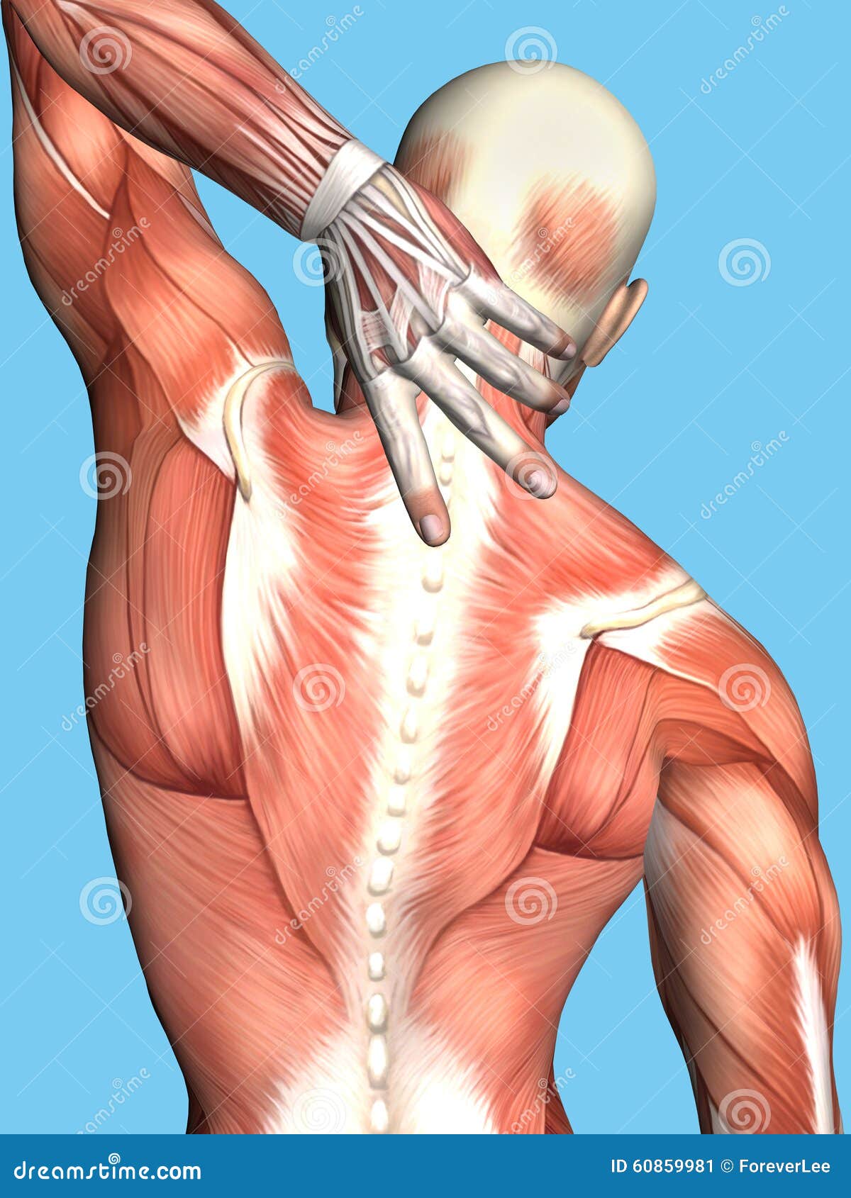
Anatomy Of Male With Upper Back Pain Stock Illustration Illustration Of Body Relief 60859981 from thumbs.dreamstime.com The bones of the chest and upper back combine to form the strong, protective rib cage around the vital thoracic organs such as the heart and lungs. See thoracic spine anatomy and upper back pain The rib cage also anchors the bones of the head, neck, shoulders, and arms to the trunk of the body. Browse 384 human anatomy organs back view stock photos and images available, or start a new search to explore more stock photos and images. Your lower back (lumbar spine) is the anatomic region between your lowest rib and the upper part of the buttock. Back muscles anatomy here include the trapezius, latissimus dorsi, rhomboid and levator scapulae. The lumbar and sacrum region make up the bone of the lower back anatomy. There is a set of muscles in the upper back (called the thoracic area) called the spinalis thoracis.
Concept or conceptual 3d human upper leg anatomy or.
It is the surface of the body opposite from the chest and the abdomen. Continue scrolling to read more below. The back consists of the spine, spinal cord, muscles, ligaments, and nerves. As viewed from the side, the cervical spine forms a lordotic curve by gently curving toward the front of the body and then back. This is my video about the muscles of the back. Human backbone diagram, bone, human backbone diagram. Female cardiovascular system, rear and front views, on black. The vertebral column runs the length of the back and creates a central area of recession. The rib cage also anchors the bones of the head, neck, shoulders, and arms to the trunk of the body. This curve, called lordosis, helps to: This muscle is located on the upper portion of the back anatomy, underneath the trapezius. The lumbar and sacrum region make up the bone of the lower back anatomy. There is a set of muscles in the upper back (called the thoracic area) called the spinalis thoracis.
Related posts of upper back muscles muscle anatomy interactive upper back anatomy. Middle part of the body.


0 Komentar